Technology has truly revolutionized our lives, and oral surgery is no exception. At Wilmington Oral Surgery, we use the latest developments in technology for maxillofacial medicine to improve the quality of care we can provide our patients.
Our 3D Cone Beam Computer Tomography (CBCT) is a perfect example of how we integrate the latest medical tech into our patient-focused oral surgery practice. Until the early 2000s, oral surgeons only had two dimensional tools at their disposal for evaluating the teeth, gums, mouth and jaw. Since we live in a 3D world, 2D imaging fell short of the precision oral and maxillofacial specialists need to offer the best care possible.
Fortunately, recent advances in dental and facial imaging enable 3D evaluations of facial and dental structures. This completely changes the game in the level of precision and detailed care we can offer, giving us a true 360 look at the intricate structures inside your mouth and face. This improves diagnostics which, in turn, improves our ability to treat any condition that affects your oral health.
In addition to increased precision, the cone beam digital technology minimizes radiation exposure compared to traditional medical CT scanners, because the machines are much smaller. But the cone beam CT still produces similar images to the scanners you would see at a hospital or outpatient clinic. It’s a healthier and more precise approach to detailed diagnostics.
Oral Surgery Tech: How the 3D Cone Beam CT Works
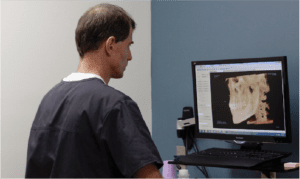
A CBCT machine requires you to place your head in a certain position so that the imager has unobstructed access to every angle of your mouth and jaw. During a scan, a focused, cone-shaped x-ray beam moves 360 degrees around your head, gathering up to 600 images, also called views. These are then integrated to produce a 3D virtual representation of your mouth.
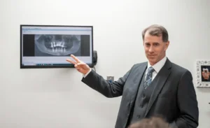
The cone beam CT allows Dr. Puckett to diagnose any issues and create a treatment plan with unmatched precision and accuracy. The system uses scatter radiation to provide clearer images than x-rays. Views and angles can be manipulated for an accurate evaluation.
Uses of CBCT Machines in Oral & Maxillofacial Medicine
CBCT has revolutionized diagnostics and treatments in numerous ways:
- Dental implant planning: Visualizes bone density and nerve pathways for precise implant placement, ensuring optimal success.
- Wisdom tooth assessment: Identifies complex wisdom tooth root anatomy and potential nerve proximity, guiding safe extraction.
- Jawbone evaluation: Accurately diagnoses jawbone fractures, cysts, and tumors, aiding treatment planning.
- Root infections/abscesses: Detects hidden infection pockets and complex root anatomy for effective treatment.
- Orthodontic planning: Analyzes jaw structure and tooth positioning for accurate treatment planning and monitoring.
- TMJ disorders: Assesses joint anatomy and movement for diagnosing and treating jaw pain.
- Facial trauma surgery: Provides detailed images of fractures for precise maxillofacial surgical interventions.
Let’s take a closer look at the three most common complications CBCT benefits patients at Wilmington Oral Surgery: dental implant planning, impacted teeth diagnoses/treatment and evaluating jaw health.
3D CBCT Optimizes Preparations for Dental Implant Surgery
Current cone beam CT technology allows for superior digital implant planning. The three-dimensional images enable superior pre-surgical treatment planning and help eliminate complications, which can occur with traditional x-rays.
Using the cone beam CT with current software, we can plan challenging implant cases that were previously considered untreatable. We can accurately assess landmarks such as the lower jawbone canal, the sinuses beside the nose and adjacent teeth, as well as the volume, height, width, and angle of bone.
The technology allows us to conduct minimally-invasive dental implant surgery, which is less painful and improves soft tissue outcomes. It also helps us optimize the placement of each dental implant, increasing chances of proper osseointegration and healing time.
Cone Beam X-rays Reveal the Details of Impacted Teeth
Additionally, the cone beam CT is an important tool to diagnose and assist with treatment of impacted teeth. Cone beam CT shows us where the impacted teeth are in relation to the roots of adjacent teeth, nerves and sinuses. These 3D views allow us to avoid damaging essential anatomical structures when the oral surgeon removes the impacted tooth.
Identifying Jaw Lesions with 3D Imaging Reduces Severity of Oral Surgeries
Three-dimensional scans help us properly determine the extent of disease in jaw lesions before our oral surgeon performs a procedure, which means we can decide the best course of treatment ahead of time. They do not always provide a specific diagnosis, but help us narrow the options and guide us in next steps.
Patient Benefits: Getting a CBCT Scan in Easy and Painless!
The scan is quick (it takes approximately ten seconds), noninvasive and painless, there are no side effects, and no radiation remains in your body. After your scan, we are able to easily share your images with your dentist, which aids collaboration on your care to provide the best outcome.
Benefits to our oral surgery patients that this advancement in oral and maxillofacial medicine has allowed us to provide include:
- Enhanced diagnosis: Reveals details invisible in traditional x-rays, leading to more accurate diagnoses and treatment plans.
- Improved treatment planning: Allows for precise planning of implant placement, surgery and other procedures.
- Minimized risk: Reduces the need for multiple x-rays, lowering radiation exposure.
- Enhanced patient experience: Shorter scan time and more comfortable positioning compared to traditional CT scans.
The Leading Edge of Technology for Oral and Maxillofacial Medicine
Part of providing the best patient care is keeping up on the latest research and developments in oral surgery tech. Our 3D Cone Beam CT allows us to diagnose and understand issues at a level of detail not possible with traditional scans. Our office also specializes in zirconia ceramic dental implants, natural post-surgery pain management and dentin bone grafting, because these new technologies assure the future of maxillofacial medicine is holistic.
Learn more about our holistic oral care services and set up a consultation with our oral surgeon for more information on how Wilmington Oral Surgery’s advanced approach to oral and maxillofacial medicine can benefit you and your family.